
Prosopagnosia (Face Blindness): Symptoms, Neurological Causes, and Diagnosis
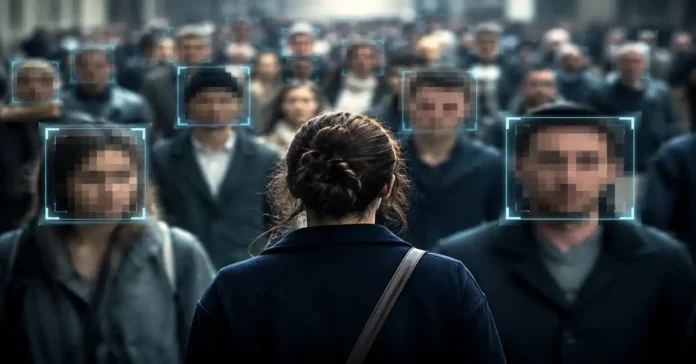
Imagine standing in a crowded grocery aisle, looking directly at your best friend, and feeling zero spark of recognition until they speak. That bewildering disconnect is daily life for someone with prosopagnosia, commonly known as face blindness. It is not an eyesight defect or general memory loss, but a specific neurocognitive breakdown in how the brain stitches visual facial geometry into a recognizable identity.
What is Prosopagnosia? symptoms and daily life
Human beings are evolutionary face specialists. Most people glance at a face and instantly register identity, age, mood, and subtle micro-expressions in less than two hundred milliseconds. For someone living with prosopagnosia, that rapid, automatic synthesis simply never happens.
Visual Input (Eyes) > Feature Breakdown (Nose, Lips) > [Processing Bottleneck] – X -> Identity Match
Prosopagnosic individuals see eyes, noses, and mouths clearly. They can tell you if a person has dark hair or thick eyebrows. However, their brain fails to assemble those individual puzzle pieces into a unified, identifiable whole — a breakdown in what cognitive psychologists call holistic processing.
Common symptoms and signs
- Failing to recognize familiar people out of context: Passing a coworker on the street or running into a neighbor at an airport without realizing who they are.
- Trouble following television shows and movies: Confusing characters who share similar hair colors or body builds.
- Relying on non-facial anchors: Identifying friends exclusively by voice tone, hairstyle, distinct jewelry, posture, or walking rhythm.
- Difficulty recognizing oneself: In severe cases, looking into a mirror or group photo and struggling to pick out one’s own reflection.
- Persistent social fatigue: Feeling ongoing anxiety in group settings where feigning recognition feels necessary to avoid seeming rude.
Because faces anchor human social bonding, people with undiagnosed prosopagnosia are frequently mislabeled as aloof, rude, or absent-minded. In reality, they are running intense mental calculations just to figure out who is saying hello.
The neural architecture: how the brain reads faces
Face recognition does not rely on a single brain switch. Instead, it depends on a coordinated neural circuit running through the ventral visual stream (the brain’s visual pathway responsible for identifying what an object is).

1. The core face network
The human brain processes faces across three interconnected stations in the cerebral cortex:
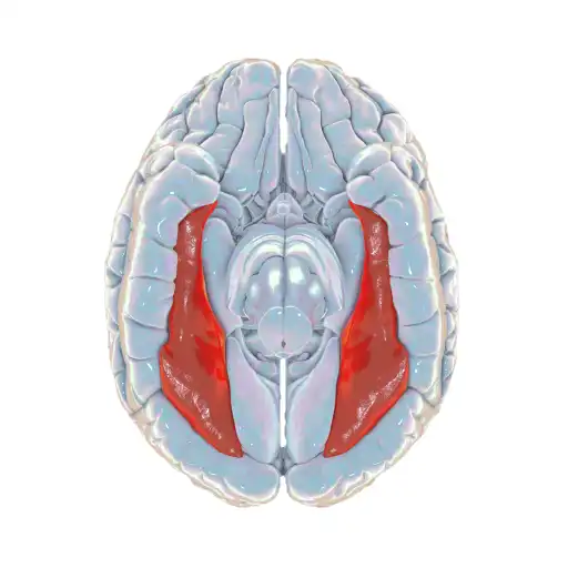
- Occipital Face Area (OFA): Located in the inferior occipital gyrus, the OFA acts as the visual gatekeeper. It registers physical facial components — calculating the curvature of an eye, the line of a jaw, or the contour of a lip.
- Fusiform Face Area (FFA): Situated on the underside of the temporal lobe within the lateral fusiform gyrus, the right FFA is the powerhouse of facial identity. It performs configural processing, mapping the exact spatial distances between facial features to create an integrated structural representation.
- Posterior Superior Temporal Sulcus (pSTS): This area tracks dynamic, moving facial signals. While the FFA handles static identity (“who is this?”), the pSTS tracks changeable features like eye gaze direction, lip movements, and shifting emotional expressions.
2. The extended network and structural bridges
Once the core network builds a visual representation, signals travel forward to the Anterior Temporal Lobe (ATL). The ATL serves as the brain’s biographical library, linking the visual image to semantic knowledge: a name, profession, personal history, and voice profile.
Information travels between these specialized hubs through dense white-matter highways, primarily the Inferior Longitudinal Fasciculus (ILF) and the Inferior Fronto-Occipital Fasciculus (IFOF). If these structural fiber tracts fail to develop properly or suffer physical damage, communication across the face-processing network falls apart.
Developmental vs Acquired Prosopagnosia
Clinicians categorize prosopagnosia based on how and when the impairment started.
| Clinical Feature | Developmental (Congenital) Prosopagnosia | Acquired Prosopagnosia |
| Onset | Present from early childhood; lifelong | Sudden onset following brain trauma or illness |
| Brain Anatomy | No visible structural lesions on standard clinical MRI scans | Detectable focal damage (strokes, tumors, traumatic injury) |
| Awareness of Deficit | Often discovered late in adolescence or adulthood | Immediate awareness; patients remember having normal face recognition |
| Prevalence | Roughly 2% to 2.5% of the general population | Rare; dependent on specific neurological injury rates |
| Underlying Mechanism | Atypical microstructural connectivity (reduced white-matter tract integrity) | Physical disruption of the FFA, OFA, or connecting neural pathways |
Apperceptive vs Associative variants
Neuropsychologists also distinguish the disorder by the specific processing stage that fails:
- Apperceptive Prosopagnosia: The perceptual breakdown happens early. The brain cannot form an accurate mental picture of the face. Individuals with this variant cannot tell whether two unfamiliar faces shown side-by-side are identical or different.
- Associative (Amnestic) Prosopagnosia: The structural perception of the face remains intact, but the brain cannot connect that image to memory. A person with associative prosopagnosia can successfully draw or match identical faces, yet the face fails to trigger any sense of familiarity or biographical identity.
Is face blindness genetic?
Yes, developmental prosopagnosia frequently runs in families.
Pedigree analyses demonstrate that hereditary prosopagnosia typically follows an autosomal dominant pattern of inheritance. That means an affected parent has a 50% chance of passing the trait to their children. Twin studies show significantly higher concordance rates for face recognition ability among monozygotic (identical) twins compared to dizygotic (fraternal) twins, establishing that facial processing efficiency is strongly heritable.

Unlike conditions tied to a single gene mutation, developmental face blindness is polygenic. It involves subtle differences during early neurodevelopment that shape how the right fusiform gyrus organizes its synaptic networks and connects with the visual cortex.
Diagnostic tests and clinical assessment
You cannot diagnose prosopagnosia with a simple eye chart or standard brain scan. Because individuals naturally develop clever compensatory habits over time, specialized neuropsychological tests strip away external clues like hair, clothing, and ears to measure facial recognition directly.
Clinical Evaluation Pipeline: Self-Report Screening (PI20) > 2. Perceptual Matching (CFPT) > 3. Short-Term Face Memory (CFMT) > 4. Semantic Recognition (Famous Faces)
1. Cambridge Face Memory Test (CFMT)
The Cambridge Face Memory Test is the gold standard for diagnosing both developmental and acquired prosopagnosia.
- How it works: Participants study several target faces cropped of hair and clothing. They are then tested across multiple stages of increasing difficulty: identifying the learned faces in different lighting, from new viewing angles, and finally beneath visual noise masks.
- Diagnostic value: Because the test uses unfamiliar faces, everyone starts with equal familiarity. A score falling two standard deviations below the population mean strongly points to a face-processing deficit.
2. Cambridge Face Perception Test (CFPT)
The CFPT examines structural sorting rather than memory. Test-takers arrange a series of morphed faces according to how closely they resemble a target face. This test helps clinicians separate apperceptive deficits (perceptual sorting errors) from associative memory deficits.
3. Famous Faces Test and the 20-Item Prosopagnosia Index (PI20)
- Famous Faces Test: Evaluates long-term recognition by presenting well-known cultural figures stripped of hair, glasses, and context.
- PI20: A validated 20-item self-report questionnaire that quantifies an individual’s everyday difficulties with facial recognition. High PI20 scores correlate reliably with objective laboratory deficits on the CFMT.
Living with Prosopagnosia: behavioral coping strategies
There is currently no pharmacological cure or surgical repair for prosopagnosia. However, individuals successfully navigate social and professional life by using deliberate, highly effective behavioral adaptations.

- Building Multimodal Profiles: Instead of waiting for a face to trigger recognition, train your focus on distinctive audio and physical traits — speech cadence, vocal pitch, unique mannerisms, posture, and gait.
- Cataloging Static Accessories: Take quick mental notes of permanent or consistent items: signature glasses frames, distinctive jewelry, tattoos, facial hair styles, or specific bags.
- Establishing Social Scripts: Keep a clear, honest explanation handy for social circles: “I have a neurological condition called face blindness. If I walk past you without waving, please don’t take it personally — say hello and tell me your name!”
- Context-Driven Anchoring: Prepare before entering gatherings. If attending a meeting, review the attendee list and note where specific colleagues plan to sit around the table.
Understanding the neurological basis of prosopagnosia helps replace self-blame with practical clarity. When you know the brain is simply processing the world through alternative channels, navigating human connection becomes far less daunting.
References
Barton, J., Corrow, S., & Dalrymple, K. (2016). Prosopagnosia: current perspectives. Eye and Brain, 8, 165–175. https://doi.org/10.2147/eb.s92838
Cited by: 215
Manippa, V., Palmisano, A., Ventura, M., & Rivolta, D. (2023). The Neural Correlates of Developmental Prosopagnosia: Twenty-Five Years on. Brain Sciences, 13(10), 1399. https://doi.org/10.3390/brainsci13101399
Cited by: 26
Rivolta, D., Woolgar, A., Palermo, R., Butko, M., Schmalzl, L., & Williams, M. A. (2014). Multi-voxel pattern analysis (MVPA) reveals abnormal fMRI activity in both the “core” and “extended” face network in congenital prosopagnosia. Frontiers in Human Neuroscience, 8. https://doi.org/10.3389/fnhum.2014.00925
Cited by: 78

